Special Neural Engineering Seminar: Harold Sackeim (Thursday, February 13 at 10am)

Title: Modern Electroconvulsive Therapy: Vastly Improved Yet Greatly Underused

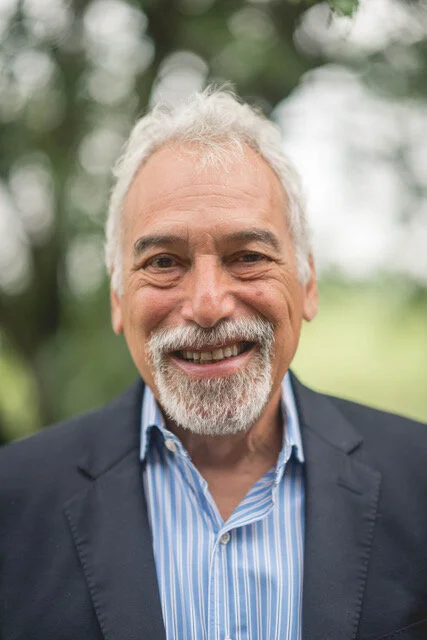
Speaker: Dr. Harold Sackeim, Professor of Psychiatry and Radiology at Columbia University and Chief of Biological Psychiatry at the New York State Psychiatric Institute

When: Thursday, February 13, 2020, 10 am to 12 noon

Where: CCNY Center for Discovery and Innovation, 3rd floor seminar room (CDI 3.352)

Contact: Marom Bikson (bikson@ccny.cuny.edu, 212-650-6791) for access to CDI building

Biography:

Dr. Harold A. Sackeim is Professor of Clinical Psychology in Psychiatry and Radiology, College of Physicians and Surgeons, Columbia University.  He served as Chief of the Department of Biological Psychiatry at the New York State Psychiatric Institute for 25 years. He is also the Founding Editor of the journal, Brain Stimulation: Basic, Translational, and Clinical Research in Neuromodulation.  He received his first B.A. from Columbia College, Columbia University (1972), another B.A. and a M.A. from Magdalen College, Oxford University (1974) and his Ph.D. from the University of Pennsylvania (1977), where he also completed his clinical training in the Department of Psychiatry. He joined the faculty of Columbia University in 1977, where he remains today.

His research has concentrated on the neurobiology and treatment of mood disorders. He has made numerous contributions to the understanding of pathophysiology of major depression and mania through use of brain imaging techniques and by examining the role of lateralization of brain function in normal emotion, neurological disorders, and psychiatric illness. For 30 years, he led the clinical research on electroconvulsive therapy (ECT) at Columbia University and the New York State Psychiatric Institute. This work has identified fundamental factors in this treatment that are responsible for its efficacy and side effects, and has radically altered understanding of both therapeutics and mechanisms of action. This research program has provided compelling evidence regarding the localization of the brain circuits involved in antidepressant effects, and has revamped understanding of the underpinnings of ECT’s effects on mood, behavior, and cognition. Dr. Sackeim is widely credited with transforming the use of this treatment worldwide.

Dr. Sackeim has directed programs at the New York State Psychiatric Institute and New York Presbyterian Hospital in the pharmacological treatment of late-life depression, and in the use of Transcranial Magnetic Stimulation (TMS), Vagus Nerve Stimulation (VNS), Deep Brain Stimulation (DBS) and other forms of focal brain stimulation. Dr. Sackeim is the inventor of Magnetic Seizure Therapy (MST), now undergoing clinical trials and has recently developed FEAST (Focal Electrically-Administered Seizure Therapy), now also in clinical trials. Dr. Sackeim introduced functional brain imaging to the medical center at Columbia in 1980, and directed a large group using Positron Emission Tomography (PET) and Magnetic Resonance Imaging (MRI) to study pathophysiology and treatment effects in mood disorders, anxiety disorders, Lyme disease, substance abuse, Alzheimer’s disease, and normal aging. Other work directed by Dr. Sackeim involved preclinical, primate research on the functional significance of structural brain changes (neurogenesis) induced by different forms of brain stimulation.

Dr. Sackeim is a member of the editorial board of several journals, and has received many national and international awards for his research contributions. These include three Distinguished Investigator Awards from the National Association for Research in Schizophrenia and Depression (NARSAD), a MERIT Award from the National Institute of Mental Health (NIMH), the Joel Elkes International Award from the American College of Neuropsychopharmacology (ACNP), election as Honorary Fellow of the American Psychiatric Association, and the Award for Research Excellence from the New York State Office of Mental Hygiene, Edward Smith Lectureship, National Institute of Psychobiology, Israel, the lifetime achievement award form the EEG and CNS Society, and the NARSAD Maddox Falcone Prize for lifetime achievement in research on affective disorders. He is past President of the Society of Biological Psychiatry and the Association for Research in Nervous and Mental Disease. He has authored more than 450 publications.
Sackeim_CCNY_Lecture_20200213.jpg
Neural Engineering
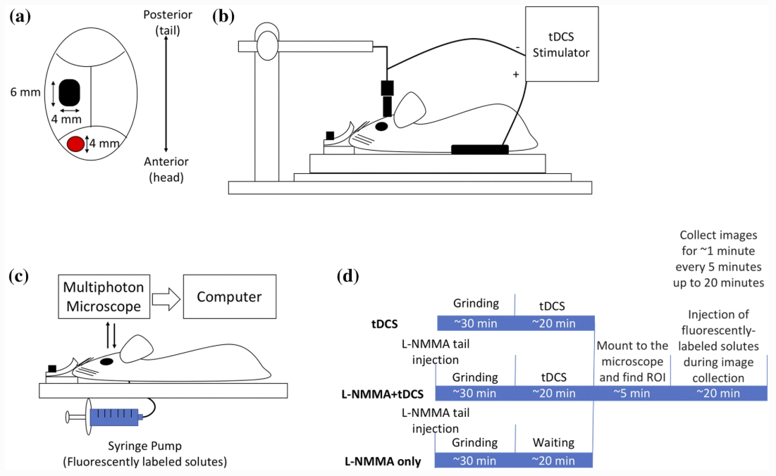
New Paper: In Vivo Modulation of the Blood–Brain Barrier Permeability by Transcranial Direct Current Stimulation (tDCS)

Shin DW, Fan J, Luu E, Khalid W, Xia Y, Khadka N, Bikson M, Fu BM. In Vivo Modulation of the Blood–Brain Barrier Permeability by Transcranial Direct Current Stimulation (tDCS). Annals of Biomedical Engineering 2020. https://doi.org/10.1007/s10439-020-02447-7


Download PDF published in Annals of Biomedical Engineering — DOI

Abstract

tDCS has been used to treat various brain disorders and its mechanism of action (MoA) was found to be neuronal polarization. Since the blood–brain barrier (BBB) tightly regulates the neuronal micro-environment, we hypothesized that another MoA of tDCS is direct vascular activation by modulating the BBB structures to increase its permeability (P). To test this hypothesis, we used high resolution multi-photon microscopy to determine P of the cerebral micro vessels in rat brain. We found that 20 min 0.1– 1 mA tDCS transiently increases P to a small solute, sodium fluorescein (MW 376) and to a large solute, Dextran-70k, with a much higher increase in P to the large solute. By pretreating the vessel with a nitric oxide synthase inhibitor, we revealed that the tDCS-induced increase in P is NO dependent. A transport model for the BBB was further employed to predict the structural changes by the tDCS. Comparing model predictions with the measured data suggests that tDCS increases P by temporarily disrupting the structural components forming the paracellular pathway of the BBB. That the transient and reversible increase in the BBB permeability also suggests new applications of tDCS such as a non-invasive approach for brain drug delivery through the BBB.

news_Article_fig.PNG
Guest User
New Video Publication: Updated Technique for Reliable, Easy, and Tolerated tES Including tDCS

Borges H, Dufau A, Paneri B, Woods AJ, Knotkova H, Bikson M. Updated Technique for Reliable, Easy, and Tolerated Transcranial Electrical Stimulation Including Transcranial Direct Current Stimulation. Journal of Visualized Experiments 2020. https://doi.org/10.3791/59204.


Download PDF published in JOVE — DOI

Neural EngineeringJOVE, tES, tDCS
New Paper: Methodology for tDCS integration with fMRI

Esmaeilpour Z, Shereen AD, Ghobadi‐Azbari P, Datta A, Woods AJ, Ironside M, O’Shea J, Kirk U, Bikson M, Ekhtiari H. Methodology for tDCS integration with fMRI. Human Brain Mapping. 2019 Dec 24; Available from: http://dx.doi.org/10.1002/hbm.24908


Download PDF published in Human Brain Mapping — DOI

Abstract

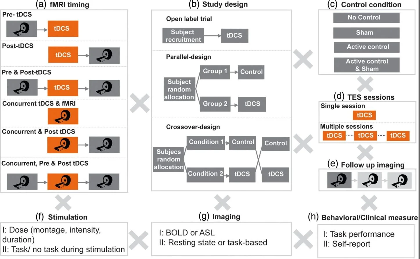
Understanding and reducing variability of response to transcranial direct current stimulation (tDCS) requires measuring what factors predetermine sensitivity to tDCS and tracking individual response to tDCS. Human trials, animal models, and computational models suggest structural traits and functional states of neural systems are the major sources of this variance. There are 118 published tDCS studies (up to October 1, 2018) that used fMRI as a proxy measure of neural activation to answer mechanistic, predictive, and localization questions about how brain activity is modulated by tDCS. FMRI can potentially contribute as: a measure of cognitive state‐level variance in baseline brain activation before tDCS; inform the design of stimulation montages that aim to target functional networks during specific tasks; and act as an outcome measure of functional response to tDCS. In this systematic review, we explore methodological parameter space of tDCS integration with fMRI spanning: (a) fMRI timing relative to tDCS (pre, post, concurrent); (b) study design (parallel, crossover); (c) control condition (sham, active control); (d) number of tDCS sessions; (e) number of follow up scans; (f) stimulation dose and combination with task; (g) functional imaging sequence (BOLD, ASL, resting); and (h) additional behavioral (cognitive, clinical) or quantitative (neurophysiological, biomarker) measurements. Existing tDCS‐fMRI literature shows little replication across these permutations; few studies used comparable study designs. Here, we use a representative sample study with both task and resting state fMRI before and after tDCS in a crossover design to discuss methodological confounds. We further outline how computational models of current flow should be combined with imaging data to understand sources of variability. Through the representative sample study, we demonstrate how modeling and imaging methodology can be integrated for individualized analysis. Finally, we discuss the importance of conducting tDCS‐fMRI with stimulation equipment certified as safe to use inside the MR scanner, and of correcting for image artifacts caused by tDCS. tDCS‐fMRI can address important questions on the functional mechanisms of tDCS action (e.g., target engagement) and has the potential to support enhancement of behavioral interventions, provided studies are designed rationally.

FIGURE 2 Methodology for tDCS integration with fMRI.jpg
Neural EngineeringtDCS, fMRI
New Paper: Experimental-design Specific Changes in Spontaneous EEG and During Intermittent Photic Stimulation by HD-tDCS

Vladimir V. Lazarev, Nigel Gebodh, Tiago Tamborino, Bikson, M, & Egas Caparelli-Daquer. (2020). Experimental-design Specific Changes in Spontaneous EEG and During Intermittent Photic Stimulation by High Definition Transcranial Direct Current Stimulation. Neuroscience, 426, 50–58. https://doi.org/10.1016/j.neuroscience.2019.11.016


Download PDF published in Journal Neuroscience Methods— DOI

Abstract

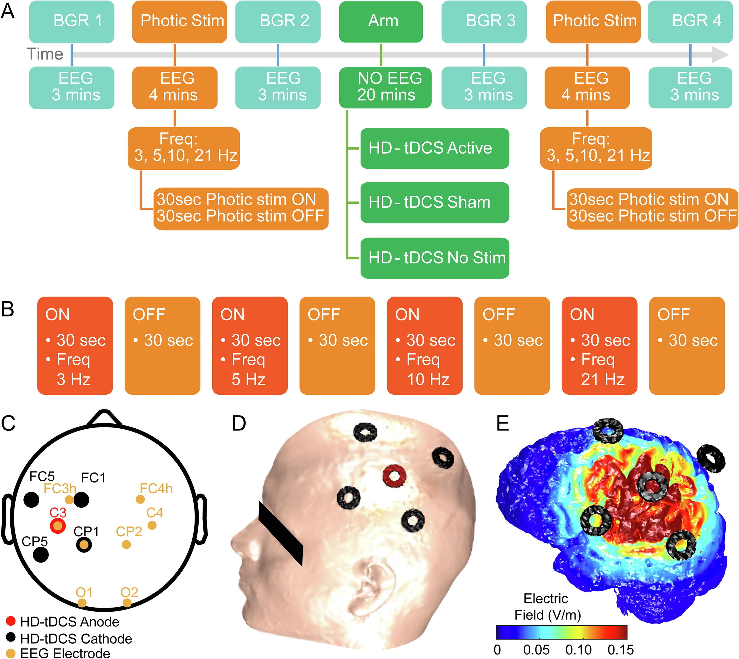
Electroencephalography (EEG) as a biomarker of neuromodulation by High Definition transcranial Direct Current Stimulation (HD-tDCS) offers promise as both techniques are deployable and can be integrated into a single head-gear. The present research addresses experimental design for separating focal EEG effect of HD-tDCS in the ‘4-cathode × 1-anode’ (4 × 1) montage over the left motor area (C3). We assessed change in offline EEG at the homologous central (C3, C4), and occipital (O1, O2) locations. Interhemispheric asymmetry was accessed for background EEG at standard frequency bands; and for the intermittent photic stimulation (IPS). EEG was compared post- vs pre-intervention in three HD-tDCS arms: Active (2 mA), Sham (ramp up/down at the start and end), and No-Stimulation (device was not powered), each intervention lasting 20 min. The asymmetric background EEG changes were only in the central areas with right-side amplitude spectra prevalence, most pronounced in the no-stimulation arm, where they depended on comparison time-points and were consistent with markers of transition between drowsiness and vigilance – bilateral decrease in the delta and asymmetric central increase in the alpha and beta1 bands. For the active arm, similar but less pronounced changes occurred in the alpha band. In contrast, responses to IPS developed similar asymmetric amplitude increase at four harmonics of the IPS of 3 Hz only in the active arm, against a background of a brain-wide symmetric increase in both active and sham arms. Our protocols and analyses suggest methodological caveats for how EEG of tDCS studies could be conducted to isolate putative brain polarization outcomes.

FIGURE 1 Experimental-design Specific Changes in Spontaneous EEG and During Intermittent Photic Stimulation by HD-tDCS.jpg
New Letter to Editor: Response to Caraway et al. on “Tissue Temperature Increases by a 10 kHz SCS System”

Niranjan Khadka, Marom Bikson. Response to the Letter to the Editor by Caraway et al. on “Tissue Temperature Increases by a 10 kHz Spinal Cord Stimulation System: Phantom and Bioheat Model”. Neuromodulation 2019. https://doi.org/10.1111/ner.13079. PDF


Download: PDF published in Neuromodulation - DOI

To the Editor:

We would like to respond to the Letter to the Editor by Dr. Caraway, Dr. Bradley, and Dr. Lee regarding our recent paper “Tissue Temperature Increases by a 10 kHz Spinal Cord Stimulation System: Phantom and Bioheat Model” 1. Caraway et al. correctly described the explicit aim of our paper “to explore the role of joule heating as a mechanism of action for HF10 therapy.” Caraway et al. also confirmed that we “conclude that the measured temperature changed in a predictable manner due to the physics of electrical heating in a volume conductor.” We then predicted temperature increases at the spinal cord and near the lead using a bioheat FEM model.

We noted in our paper that “the bioheat models of kHz SCS remain to be validated” and Caraway et al. emphasized that we did not include in vivo (preclinical or clinical) data. In this paper, and our prior publication on the general topic “Temperature Increases by kilohertz frequency Spinal Cord Stimulation” 2, we explicitly considered how computational models' assumption may increase or decrease predicted temperature rises. If ongoing validation confirms heating of ~1°C, there are key outstanding questions on if and how pain processing pathways are affected. In the spirit of “meritorious scientific discussion,” we may differ with Caraway et al. on the potential impact of moderate heating as a mechanism of action and how this supports findings from clinical trials. But, we agree it is a misinterpretation of our work to suggest a prediction of ~1°C heating is evidence disproving preclinical or clinical data on the safety of HF10.

ner12980-fig-0003-m.jpg
Guest User
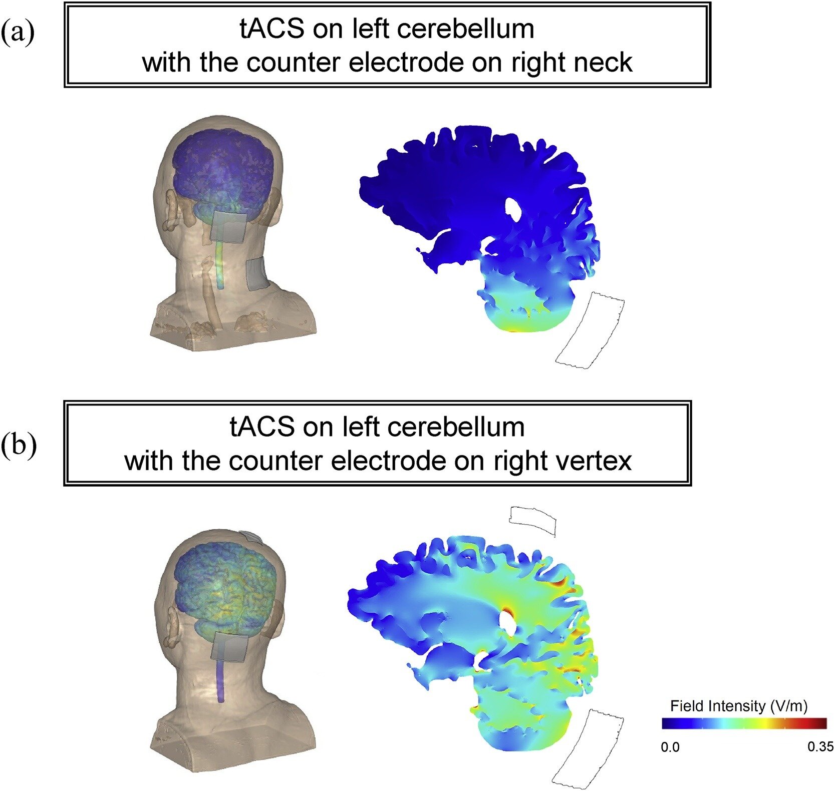
New Paper: Cerebellar tACS modulates human gait rhythm

Koganemaru S, Mikami Y, Matsuhashi M, Truong DQ, Bikson M, Kansaku K, Mima T. Cerebellar transcranial alternating current stimulation modulates human gait rhythm. Neuroscience Research. 2019 Dec; Available from: http://dx.doi.org/10.1016/j.neures.2019.12.003


Download PDF published in Neuroscience Research — DOI

Abstract

Although specific brain regions are important for regularly patterned limb movements, the rhythm generation system that governs bipedal locomotion in humans is not thoroughly understood. We investigated whether rhythmic transcranial brain stimulation over the cerebellum could alter walking rhythm. Fourteen healthy subjects performed over-ground walking for 10 min during which they were given, in a random order, transcranial alternating current stimulation (tACS) over the left cerebellum at the approximated frequency of their gait cycle, tACS over the skin of the scalp, and during sham stimulation. Cerebellar tACS showed a significant entrainment of gait rhythm compared with the control conditions. When the direction of the tACS currents was symmetrically inverted, some subjects showed entrainment at an approximately 180° inverted phase, suggesting that gait modulation is dependent on current orientation. These findings indicate that tACS over cerebellum can modulate gait generation system in cerebellum and become an innovative approach for the recovery of locomotion in patients with gait disturbances caused by CNS disorders.

Figure 4 Cerebellar tACS modulates human gait rhythm.jpg
New PrePrint: Realistic Anatomically Detailed Open-Source Spinal Cord Stimulation (RADO-SCS) Model

Niranjan Khadka, Xijie Liu, Hans Zander, Jaiti Swami, Evan Rogers, Scott F. Lempka, Marom Bikson. Realistic Anatomically Detailed Open-Source Spinal Cord Stimulation (RADO-SCS) Model. bioRxiv. 2019. https://doi.org/10.1101/857946


Download: PDF published in bioRxiv — DOI

Abstract

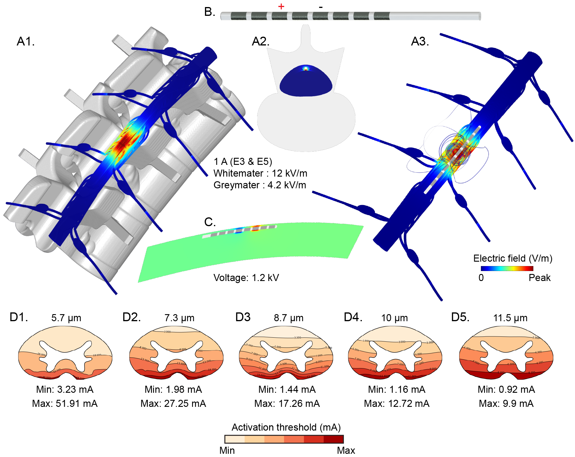
Objective: Computational current flow models of spinal cord stimulation (SCS) are widely used in device development, clinical trial design, and patient programming. Proprietary models of varied sophistication have been developed. An open-source model with state-of-the-art precision would serve as a standard for SCS simulation.

Approach: We developed a sophisticated SCS modeling platform, named Realistic Anatomically Detailed Open-Source Spinal Cord Stimulation (RADO-SCS) model. This platform consists of realistic and detailed spinal cord and ancillary tissues anatomy derived based on prior imaging and cadaveric studies. Represented tissues within the T9-T11 spine levels include vertebrae, intravertebral discs, epidural space, dura, CSF, white-matter, gray-matter, dorsal and ventral roots and rootlets, dorsal root ganglion, sympathetic chain, thoracic aorta, epidural space vasculature, white-matter vasculature, and thorax. As an exemplary, a bipolar SCS montage was simulated to illustrate the model workflow from the electric field calculated from a finite element model (FEM) to activation thresholds predicted for individual axons populating the spinal cord.

Main Results: Compared to prior models, RADO-SCS meets or exceeds detail for every tissue compartment. The resulting electric fields in white and gray-matter, and axon model activation thresholds are broadly consistent with prior stimulations.

Significance: The RADO-SCS can be used to simulate any SCS approach with both unprecedented resolution (precision) and transparency (reproducibility). Freely available online, the RADO-SCS will be updated continuously with version control.

fig2_prediciton.png
Guest User
Bikson speaks at Cleveland FES Center, Dec 5 (live Webinar)

Update watch talk video here

Prof. Marom Bikson speaks at the Cleveland FES Center NEURAL PROTHESIS LIVE WEBINAR on Dec 5, 2019 at 3:00 PM. Live Stream at: http://fescenter.org/news-events/webinar-series/

Download presentation slides

Title: Neuromodulation though BBB stimulation or Heating: New Mechanisms of DBS, SCS, and tDCS

Abstract: This seminar explores when direct heating of tissue or direct stimulation of endothelial cells of the blood-brain barrier (BBB) is the the mechanisms of action in neuromodulation. Specific approaches considered are Spinal Cord Stimulation (SCS), kHz-frequency SCS, Deep Brain Stimulation (DBS), kHz-frequency DBS, and transcranial Direct Current Stimulation.

Results are based on computational modeling (including bio-heat FEM), phantoms, and animal models (including in vivo imaging, brain slices, and in vitro endothelial monolayers). Moderate (~1 C) non-injurious heating of tissue results from higher-power waveforms with implanted electrodes (kHZ SCS, high-density SCS, kHZ DBS). Especially when sustained over long periods, moderate heating will modulate brain function. Immediate and lasting non-injurious changes in BBB permeability can occur during SCS, DBS, and tDCS. Even small changes in BBB function can have profound effects on brain function. These results are shown to derive from well established physical principles such as joule heat and electroosmosis.

In some cases heating or BBB mechanisms operate parallel to traditional nervous tissue stimulation, and in others applications they may underpin responsiveness and so govern secondary neuronal changes.

DownloadL Flier

Screen Shot 2019-11-25 at 2.57.24 PM.png
Screen Shot 2019-11-25 at 2.55.47 PM.png
Marom Bikson
New Paper: Beyond the target area: an integrative view of tDCS-induced motor cortex modulation in patients and athletes

Morya E, Monte-Silva K, Bikson M, Esmaeilpour Z, Biazoli CE, Fonseca A, Bocci T, Farzan F, Chatterjee R, Hausdorff JM, da Silva Machado DG, Russowsky Brunoni A, Mezger E, Aparecida Mascaleski L, Pegado R, Sato JR, Caetano MS, Nunes Sá K, Tanaka C, Li LM, Fontes Baptista A, Hideki Okano A. Beyond the target area: an integrative view of tDCS-induced motor cortex modulation in patients and athletes. Journal of NeuroEngineering and Rehabilitation. 2019 Nov 15;16(1). Available from: http://dx.doi.org/10.1186/s12984-019-0581-1


Download PDF published in Journal of NeuroEngineering and Rehabilitation — DOI

Abstract

Transcranial Direct Current Stimulation (tDCS) is a non-invasive technique used to modulate neural tissue. Neuromodulation apparently improves cognitive functions in several neurologic diseases treatment and sports performance. In this study, we present a comprehensive, integrative review of tDCS for motor rehabilitation and motor learning in healthy individuals, athletes and multiple neurologic and neuropsychiatric conditions. We also report on neuromodulation mechanisms, main applications, current knowledge including areas such as language, embodied cognition, functional and social aspects, and future directions. We present the use and perspectives of new developments in tDCS technology, namely high-definition tDCS (HD-tDCS) which promises to overcome one of the main tDCS limitation (i.e., low focality) and its application for neurological disease, pain relief, and motor learning/rehabilitation. Finally, we provided information regarding the Transcutaneous Spinal Direct Current Stimulation (tsDCS) in clinical applications, Cerebellar tDCS (ctDCS) and its influence on motor learning, and TMS combined with electroencephalography (EEG) as a tool to evaluate tDCS effects on brain function.

Figure 1 Beyond the target area an integrative view of tDCS-induced motor cortex modulation in patients and athletes.JPG
New Paper: Electric field causes volumetric changes in the human brain

Argyelan M, Oltedal L, Deng Z-D, Wade B, Bikson M, Joanlanne A, Sanghani S, Bartsch H, Cano M, Dale AM, Dannlowski U, Dols A, Enneking V, Espinoza R, Kessler U, Narr KL, Oedegaard KJ, Oudega ML, Redlich R, Stek ML, Takamiya A, Emsell L, Bouckaert F, Sienaert P, Pujol J, Tendolkar I, van Eijndhoven P, Petrides G, Malhotra AK, Abbott C. Electric field causes volumetric changes in the human brain. eLife. 2019 Oct 23;8. Available from: http://dx.doi.org/10.7554/eLife.49115


Download PDF published in eLife — DOI

Abstract

Recent longitudinal neuroimaging studies in patients with electroconvulsive therapy (ECT) suggest local effects of electric stimulation (lateralized) occur in tandem with global seizure activity (generalized). We used electric field (EF) modeling in 151 ECT treated patients with depression to determine the regional relationships between EF, unbiased longitudinal volume change, and antidepressant response across 85 brain regions. The majority of regional volumes increased significantly, and volumetric changes correlated with regional electric field (t = 3.77, df = 83, r = 0.38, p=0.0003). After controlling for nuisance variables (age, treatment number, and study site), we identified two regions (left amygdala and left hippocampus) with a strong relationship between EF and volume change (FDR corrected p<0.01). However, neither structural volume changes nor electric field was associated with antidepressant response. In summary, we showed that high electrical fields are strongly associated with robust volume changes in a dose-dependent fashion.

Figure 1  Electric field causes volumetric changes in the human brain.jpg
Prof. Bikson featured on Behind the Bench
Screen Shot 2019-10-17 at 11.18.37 AM.png

Prof. Marom Bikson interviewed for Behind the Bench. Includes discussion of how the the Bikson’s lab unique approach to neural engineering and medical device creation.

“There was not much work at CWRU in non-invasive stimulation. By picking that area for my research I had to leverage a lot of the tools that were developed at Case, but I also had to adapt them and create a new sort of toolkit of neural engineering for non-invasive electrical stimulation,” Bikson said. “I’m starting to work more and more with them,” he continued. “For example, I’m working with Rafael Carbunaru, who was a student of Dominique’s and now he’s directing R&D at Boston Scientific. And the reason these things came full circle is because all of a sudden, the fields of invasive and non-invasive neuromodulation that were seemingly different started to push in against each other. All of a sudden, high frequency became relevant for non-invasive and all of a sudden, sub-threshold became relevant for invasive. And oscillations became relevant for everything. Closed loop became relevant for everything. And in this way, I’m starting to work more in spinal cord stimulation, but I’m doing it with toolkits that we developed for non-invasive neuromodulation. Another example is individualized modeling was pioneered first for invasive but was pushed ahead for non-invasive. A lot of those tools can now be applied back to invasive.”

Read the full article: https://www.neurotechbench.com/post/marom-bikson-is-making-an-impact

Marom Bikson
New Paper: DCS boosts hebbian plasticity in vitro

Kronberg G, Rahman A, Sharma M, Bikson M, Parra LC. Direct current stimulation boosts hebbian plasticity in vitro. Brain Stimulation. 2019 Oct; Available from: http://dx.doi.org/10.1016/j.brs.2019.10.014


Download PDF published in Brain Stimulation — DOI

Abstract

There is evidence that transcranial direct current stimulation (tDCS) can improve learning performance. Arguably, this effect is related to long term potentiation (LTP), but the precise biophysical mechanisms remain unknown. We propose that direct current stimulation (DCS) causes small changes in postsynaptic membrane potential during ongoing endogenous synaptic activity. The altered voltage dynamics in the postsynaptic neuron then modify synaptic strength via the machinery of endogenous voltage-dependent Hebbian plasticity. This hypothesis predicts that DCS should exhibit Hebbian properties, namely pathway specificity and associativity. We studied the effects of DCS applied during the induction of LTP in the CA1 region of rat hippocampal slices and using a biophysical computational model. DCS enhanced LTP, but only at synapses that were undergoing plasticity, confirming that DCS respects Hebbian pathway specificity. When different synaptic pathways cooperated to produce LTP, DCS enhanced this cooperation, boosting Hebbian associativity. Further slice experiments and computer simulations support a model where polarization of postsynaptic pyramidal neurons drives these plasticity effects through endogenous Hebbian mechanisms. The model is able to reconcile several experimental results by capturing the complex interaction between the induced electric field, neuron morphology, and endogenous neural activity. These results suggest that tDCS can enhance associative learning. We propose that clinical tDCS should be applied during tasks that induce Hebbian plasticity to harness this phenomenon, and that the effects should be task specific through their interaction with endogenous plasticity mechanisms. Models that incorporate brain state and plasticity mechanisms may help to improve prediction of tDCS outcomes.

FIGURE 1 Direct current stimulation boosts hebbian plasticity in vitro.jpg
New Paper: The Quasi-uniform assumption for Spinal Cord Stimulation translational research

Khadka N, Truong DQ, Williams P, Martin JH, Bikson M. The Quasi-uniform assumption for Spinal Cord Stimulation translational research. Journal of Neuroscience Methods. 201. ;328:108446. http://dx.doi.org/10.1016/j.jneumeth.2019.108446


Download PDF published in Journal Neuroscience Methods— DOI

Abstract

Background: Quasi-uniform assumption is a general theory that postulates local electric field predicts neuronal activation. Computational current flow model of spinal cord stimulation (SCS) of humans and animal models inform how the quasi-uniform assumption can support scaling neuromodulation dose between humans and translational animal.

New method: Here we developed finite element models of cat and rat SCS, and brain slice, alongside SCS models. Boundary conditions related to species specific electrode dimensions applied, and electric fields per unit current (mA) predicted.

Results: Clinically and across animal, electric fields change abruptly over small distance compared to the neuronal morphology, such that each neuron is exposed to multiple electric fields. Per unit current, electric fields generally decrease with body mass, but not necessarily and proportionally across tissues. Peak electric field in dorsal column rat and cat were ∼17x and ∼1x of clinical values, for scaled electrodes and equal current. Within the spinal cord, the electric field for rat, cat, and human decreased to 50% of peak value caudo-rostrally (C5–C6) at 0.48 mm, 3.2 mm, and 8 mm, and mediolaterally at 0.14 mm, 2.3 mm, and 3.1 mm. Because these space constants are different, electric field across species cannot be matched without selecting a region of interest (ROI)

Comparison of existing method: This is the first computational model to support scaling neuromodulation dose between humans and translational animal.

Conclusions: Inter-species reproduction of the electric field profile across the entire surface of neuron populations is intractable. Approximating quasi-uniform electric field in a ROI is a rational step to translational scaling.

graphical_abstract.jpg
Guest User
CCNY Neural Engineering lab presents innovative projects at 2019 Meeting of Neuromodulation: The Science & NYC Neuromodulation
  1. Can kilohertz-frequency Deep Brain Stimulation increase brain tissue temperature?

    Niranjan Khadka, Irene Harmsen, Andres M. Lozano, Marom Bikson

  2. Waveform Characterization, IPG battery life, and Temperature Increases by Senza HF10 Spinal Cord Stimulation System

    Adantachede L Zannou, Niranjan Khadka, Mohmad FallahRad, Dennis Q. Truong, Brian H Kopell, Marom Bikson

  3. Modulation of Sleepiness and Physiology with Brain-Derived and Narrow-Band tACS

    Nigel Gebodh, Laura Vacchi, Zeinab Esmaeilpour, Devin Adair, Alexander Poltorak, Valeria Poltorak, Marom Bikson

  4. Realistic Anatomically Detailed Open-source Spinal Cord Stimulation (RADO SCS) Model

    Niranjan Khadka, Xijie Liu, Jaiti Swami, Hans Zander, Evan Rogers, Scott Lempka, Marom Bikson

    Download: PDF

  5. Mechanism of Temporal Interference (TI) stimulation

    Zeinab Esmaeilpour, Greg Kronberg, Lucas Parra, Marom Bikson

And Prof. Marom Bikson chairs and presents at Session 1: Session 1: New Engineering of Neuromodulation & Brain Machine Interfaces on “Personalized Neuromodulation: Reading the Brain to Write the Brain.” Download Slide: PDF

Embed Block
Add an embed URL or code. Learn more
Embed Block
Add an embed URL or code. Learn more
Guest User
New Paper: CNS Electrical Stimulation for Neuroprotection in Acute Cerebral Ischemia

Bahr Hosseini M, Hou J, Bikson M, Iacoboni M, Gornbein J, Saver JL. Central Nervous System Electrical Stimulation for Neuroprotection in Acute Cerebral Ischemia. Stroke. 2019 Oct;50(10):2892–901. https://doi.org/10.1161/STROKEAHA.119.025364.


Download PDF published in Stroke — DOI

Abstract

Brain electrical stimulation, widely studied to facilitate recovery from stroke, has also been reported to confer direct neuroprotection in preclinical models of acute cerebral ischemia. Systematic review of controlled preclinical acute cerebral ischemia studies would aid in planning for initial human clinical trials. A systematic Medline search identified controlled, preclinical studies of central nervous system electrical stimulation in acute cerebral ischemia. Studies were categorized among 6 stimulation strategies. Three strategies applied different stimulation types to tissues within the ischemic zone (cathodal hemispheric stimulation [CHS], anodal hemispheric stimulation, and pulsed hemispheric stimulation), and 3 strategies applied deep brain stimulation to different neuronal targets remote from the ischemic zone (fastigial nucleus stimulation, subthalamic vasodilator area stimulation, and dorsal periaqueductal gray stimulation). Random-effects meta-analysis assessed electrical stimulation modification of final infarct volume. Study-level risk of bias and intervention-level readiness-for-translation were assessed using formal rating scales. Systematic search identified 28 experiments in 21 studies, including a total of 350 animals, of electrical stimulation in preclinical acute cerebral ischemia. Overall, in animals undergoing electrical stimulation, final infarct volumes were reduced by 37% (95% CI, 34%–40%; P<0.001), compared with control. There was evidence of heterogeneity of efficacy among stimulation strategies (I2=93.1%, P_heterogeneity<0.001). Among the within-ischemic zone stimulation strategies, only CHS significantly reduced the infarct volume (27 %; 95% CI, 22%–33%; P<0.001); among the remote-from ischemic zone approaches, all (fastigial nucleus stimulation, subthalamic vasodilator area stimulation, and dorsal periaqueductal gray stimulation) reduced infarct volumes by approximately half. On formal rating scales, CHS studies had the lowest risk of bias, and CHS had the highest overall quality of intervention-level evidence supporting readiness to proceed to clinical testing. Electrical stimulation reduces final infarct volume across preclinical studies. CHS shows the most robust evidence and is potentially appropriate for progression to early-stage human clinical trial testing as a promising neuroprotective intervention.

FIGURE 1 CNS Electrical Stimulation for Neuroprotection in Acute Cerebral Ischemia.JPG
New Paper: Adaptive current tDCS up to 4 mA

Niranjan Khadka, Helen Borges, Bhaskar Paneri, Trynia Kaufman, Electra Nassis, Adantchede L. Zannou, Yungjae Shin, Hyeongseob Choi, Seonghoon Kim, Kiwon Lee, Marom Bikson. Adaptive current tDCS up to 4 mA. Brain Stimulation. 2019 Jan;13(1):69–79. https://doi.org/10.1016/j.brs.2019.07.027


Download: PDF published in Brain Stimulation — DOI

Abstract

Background: Higher tDCS current may putatively enhance efficacy, with tolerability the perceived limiting factor.

Objective: We designed and validated electrodes and an adaptive controller to provide tDCS up to 4 mA,while managing tolerability. The adaptive 4 mA controller included incremental ramp up, impedance-based current limits, and a Relax-mode where current is transiently decreased. Relax-mode was automatically activated by self-report VAS-pain score>5 and in some conditions by a Relax-button available to participants.

Methods: In a parallel-group participant-blind design with 50 healthy subjects, we used specialized electrodes to administer 3 daily session of tDCS for 11 min, with a lexical decision task as a distractor, in 5 study conditions: adaptive 4 mA, adaptive 4 mA with Relax-button, adaptive 4 mA with historical-Relax-button, 2 mA, and sham. A tablet-based stimulator with a participant interface regularly queried VAS pain score and also limited current based on impedance and tolerability. An Abort-button provided in all conditions stopped stimulation. In the adaptive 4 mA with Relax-button and adaptive 4 mA with historical-Relax-button conditions, participants could trigger a Relax-mode ad libitum, in the latter case with incrementally longer current reductions. Primary outcome was the average current delivered during each session, VAS pain score, and adverse event questionnaires. Current delivered was analyzed either excluding or including dropouts who activated Abort (scored as 0 current).

Results: There were two dropouts each in the adaptive 4 mA and sham conditions. Resistance based current attenuation was rarely activated, with few automatic VAS pain score triggered relax-modes. In conditions with Relax-button option, there were significant activation often irrespective of VAS pain score. Including dropouts, current across conditions were significantly different from each other with maximum current delivered during adaptive 4 mA with Relax-button. Excluding dropouts, maximum current was delivered with adaptive 4 mA. VAS pain score and adverse events for the sham was only significantly lower than the adaptive 4 mA with Relax-button and adaptive 4 mA with historical-Relax-button. There was no difference in VAS pain score or adverse events between 2 mA and adaptive 4 mA.

Conclusions: Provided specific electrodes and controllers, adaptive 4 mA tDCS is tolerated and effectively blinded, with acceptability likely higher in a clinical population and absence of regular querying. Indeed,presenting participants with overt controls increases rumination on sensation.

Fig_1.jpg
Bikson features in IEEE "How a Tiny Electrical Current to the Brain is Treating Medical Symptoms"
Screen Shot 2019-08-06 at 12.47.55 PM.png

Prof. Marom Bikson discusses both home-based tDCS and HD-tDCS in IEEE “How a Tiny Electrical Current to the Brain Is Treating Medical Symptoms”.

Read the article: PDF

“Bikson, professor of biomedical engineering at City University of New York (CUNY). He is also cofounder of Soterix Medical of New York City, which is developing tDCS for home, clinical, and research applications. On the medical-supervision side, Soterix is working on an automated home system that a clinician (or researcher if it’s part of a research project) can monitor and direct via telemedicine (Figure 4). “There’s a lot of technology that goes into that, including reliable communication between the technology and the site that’s providing the telemedicine, as well as automated software that can collect information from the patient on a daily basis, and can also deliver the prescription that the doctor sets,” Bikson said. “Although the software doesn’t decide what the treatment will be, it can withhold treatment until the patient is ready to receive it based on the doctor’s prescription schedule.”

“Going high-def: Besides the home-based tDCS system, Soterix also developed something quite different for use in research labs and clinics: targeted tDCS that delivers current to precise, small areas of the brain, as compared to the 5x5-cm areas covered by each of the two pads or sponges seen in typical home based systems, Bikson explained. The idea with targeted tDCS is to personalize treatment for individual patients and/or to reduce distinct symptoms. Soterix actually engineered the first noninvasive, targeted, and low-intensity neuromodulation technology, dubbed high-definition tDCS (HD-tDCS), back in 2009. “HD-tDCS and its variants, which include HD-tACS, use an array of smaller electrodes to stimulate specific parts of the brain,”

Screen Shot 2019-08-06 at 12.53.51 PM.png
Marom Bikson