New Paper: Intracranial voltage recording during transcranial direct current stimulation (tDCS) in human subjects with validation of a standard model

Esmaeilpour Z, Milosevic M, Azevedo K, Khadka N, Navarro J, Brunoni A, Popovic MR, Bikson M, Fonoff ET


Download: PDF published in Brain Stimulation  DOI

Abstract

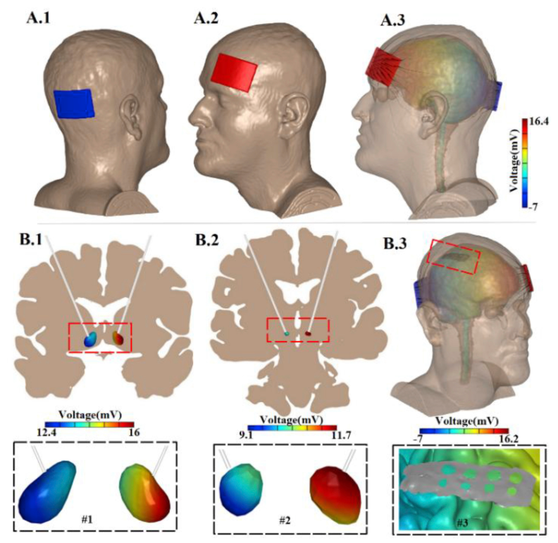
During transcranial direct current stimulation (tDCS) weak (1-2 mA) currents are applied across the head, producing low-intensity electric fields in the brain with the intention of modulating neuronal function. For any application of tDCS spanning cognitive neuroscience and neuropsychiatric therapies [1], understanding the amount of current delivered to the brain and the resulting electric field (in V/m) produced is thus important. In animal studies, direct current (DC) electric fields as low as 0.2-1.0 V/m influence neuronal excitability and plasticity [2, 3]. Since measurement of electric field in human is difficult to implement, high-resolution finite element head models [4] have been used to predict brain current flow during tDCS [5] – with many reports adapting a standard (S#) head [6-8]. There have been previous attempts to validate computational model predictions indirectly with scalp electrodes [9] and neurophysiology [10] during tDCS, as well as directly using intra-cranial electrodes, but not with DC stimulation [11, 12]. In this pilot study, DC voltage was measured using deep brain stimulation (DBS) and epidural lead electrodes during application of tDCS in human subjects. The results were evaluated against a standard (S#) head model. The model predictions of voltage produced across cortical (epidural) electrodes were consistent with recorded data, while subcortical (DBS) voltages were sensitive to conductivity assigned to subcortical structures.

12-20-17 image.PNG
Neural Engineering
New Paper: Motor cortex and spinal cord neuromodulation promote corticospinal tract axonal outgrowth and motor recovery after cervical contusion spinal cord injury

N. Zareen , M. Shinozaki , D. Ryan , H. Alexander , A. Amer, D.Q. Truong, N. Khadka, A. Sarkar, S. Naeem, M. Bikson , J.H. Martin


Download: PDF published in  Experimental Neurology DOI

Abstract

Cervical injuries are the most common form of SCI. In this study, we used a neuromodulatory approach to promote skilled movement recovery and repair of the corticospinal tract (CST) after a moderately severe C4 midline contusion in adult rats. We used bilateral epidural intermittent theta burst (iTBS) electrical stimulation of motor cortex to promote CST axonal sprouting and cathodal trans-spinal direct current stimulation (tsDCS) to enhance spinal cord activation to motor cortex stimulation after injury. We used Finite Element Method (FEM) modeling to direct tsDCS to the cervical enlargement. Combined iTBS-tsDCS was delivered for 30 min daily for 10 days. We compared the effect of stimulation on performance in the horizontal ladder and the Irvine Beattie and Bresnahan forepaw manipulation tasks and CST axonal sprouting in injury-only and injury + stimulation animals. The contusion eliminated the dorsal CST in all animals. tsDCS significantly enhanced motor cortex evoked responses after C4 injury. Using this combined spinal-M1 neuromodulatory approach, we found significant recovery of skilled locomotion and forepaw manipulation skills compared with injury-only controls. The spared CST axons caudal to the lesion in both animal groups derived mostly from lateral CST axons that populated the contralateral intermediate zone. Stimulation enhanced injury-dependent CST axonal outgrowth below and above the level of the injury. This dual neuromodulatory approach produced partial recovery of skilled motor behaviors that normally require integration of postureupper limb sensory information, and intent for performance. We propose that the motor systems use these new CST projections to control movements better after injury.

12-20-17 image 2.PNG
Neural Engineering
New Paper: Incomplete evidence that increasing current intensity of tDCS boosts outcomes

New Paper: Incomplete evidence that increasing current intensity of tDCS boosts outcomes

Download: PDF published in Brain Stimulation – doi.org/10.1016/j.brs.2017.12.002

Zeinab Esmaeilpour, Paola Marangolo, Benjamin M. Hampstead, Sven Bestmann, Elisabeth Galletta, Helena Knotkova, Marom Bikson


ABSTRACT
Background: Transcranial direct current stimulation (tDCS) is investigated to modulate neuronal function by applying a fixed low-intensity direct current to scalp.

Objectives: We critically discuss evidence for a monotonic response in effect size with increasing current intensity, with a specific focus on a question if increasing applied current enhance the efficacy of tDCS.

Methods: We analyzed tDCS intensity does-response from different perspectives including biophysical modeling, animal modeling, human neurophysiology, neuroimaging and behavioral/clinical measures. Further, we discuss approaches to design dose-response trials.

Results: Physical models predict electric field in the brain increases with applied tDCS intensity. Data from animal studies are lacking since a range of relevant low-intensities is rarely tested. Results from imaging studies are ambiguous while human neurophysiology, including using transcranial magnetic stimulation (TMS) as a probe, suggests a complex state-dependent non-monotonic dose response. The diffusivity of brain current flow produced by conventional tDCS montages complicates this analysis, with relatively few studies on focal High Definition (HD)-tDCS. In behavioral and clinical trials, only a limited range of intensities (1-2 mA), and typically just one intensity, are conventionally tested; moreover, outcomes are subject brain-state dependent. Measurements and models of current flow show that for the same applied current, substantial differences in brain current occur across individuals. Trials are thus subject to inter-individual differences that complicate consideration of population-level dose response.

Conclusion: The presence or absence of simple dose response does not impact how efficacious a given tDCS dose is for a given indication. Understanding dose-response in human applications of tDCS is needed for protocol optimization including individualized dose to reduce outcome variability, which requires intelligent design of dose-response studies.

12-8-17 image.PNG
Neural Engineering
Neural Engineering Group hosts workshop on Deep Learning Methods of EEG

A Short Practicum on Deep Learning for EEG-based Brain Computer Interfaces
Presented by Vernon Lawhern of the Army Research Laboratory
Location: CDI 1.352 (First floor conference room)
Date and Time: Thursday, November 30, 2017 from 11am to 2pm
Lunch will be provided! Bring a laptop with Python Installed

Vernon will be available on Thursday afternoon and Friday morning for those who would like to try and obtain feedback on methods outlined in the workshop

See flyer below on how to RSVP and for more information

11-30-17 image 1.PNG
Neural Engineering
Dr. Bikson in US News and World Report, Nov 17, 2017

Dr. Marom Bikson interviewed by US News and World Report for a feature on

Can Transcranial Stimulation Help With Depression?

“Brain zapping”  helps patients who don’t respond to other treatments.

Read it link

“Bikson sees great strides being made in the coming years. “We are at baby aspirin levels of dosage and flip-phone levels of technology,” he says. “We have not even scratched the surface. We haven’t seen anything yet in the potential of electroceuticals.”

Neural Engineering
New Paper: High-Definition transcranial direct current stimulation in early onset epileptic encephalopathy: a case study

New Paper: High-Definition transcranial direct current stimulation in early onset epileptic encephalopathy: a case study

Download: PDF published in Brain Injury doi.org/10.1080/02699052.2017.1390254

Oded Meiron, Rena Gale, Julia Namestnic, Odeya Bennet-Back, Jonathan Davia, Nigel Gebodh, Devin Adair, Zeinab Esmaeilpour, and Marom Bikson

ABSTRACT

Primary objective: Early onset epileptic encephalopathy is characterized by high daily seizure-frequency, multifocal epileptic discharges, severe psychomotor retardation, and death at infancy. Currently, there are no effective treatments to alleviate seizure frequency and high-voltage epileptic discharges in these catastrophic epilepsy cases. The current study examined the safety and feasibility of High-Definition transcranial direct current stimulation (HD-tDCS) in reducing epileptiform activity in a 30-month-old child suffering from early onset epileptic encephalopathy.

Design and Methods: HD-tDCS was administered over 10 intervention days spanning two weeks including pre- and post-intervention video-EEG monitoring. Results: There were no serious adverse events or side effects related to the HD-tDCS intervention. Frequency of clinical seizures was not significantly reduced. However, interictal sharp wave amplitudes were significantly lower during the post-intervention period versus baseline. Vital signs and blood biochemistry remained stable throughout the entire study.

Conclusions: These exploratory findings support the safety and feasibility of 4 × 1 HD-tDCS in early onset epileptic encephalopathy and provide the first evidence of HD-tDCS effects on paroxysmal EEG features in electroclinical cases under the age of 36 months. Extending HD-tDCS treatment may enhance electrographic findings and clinical effects.

11-20-17 image2.PNG
Neural Engineering
Neural Engineering Journal Club (Thursday Nov. 16)

Event Time and Location: Thursday 11/16 at noon in CDI 3.352

Zeinab Esmaeilpour will present her work on brain stimulation and fMRI

tDCS integration with functional magnetic resonance imaging (fMRI)

Abstract:

Transcranial direct current stimulation (tDCS) is a non-invasive stimulation method that provides clinicians and researchers a tool to modulate central nervous system excitability in human and thereby contribute to exploration of brain-behavior relationship and develop treatment for various neurological and psychiatric disorders. However, despite its obvious promise, the potential of tDCS cannot be fully exploited as there is still a lack of understanding of the neural mechanisms underpinning stimulation. A key methodological advance toward bridging the gap in our understanding of the neural mechanisms of tDCS effects involves integration of tDCS with modern clinical and cognitive neuroscience techniques.

Magnetic resonance imaging (MRI) provides a high degree of spatial resolution regarding both brain structure and function, with the ability to assess brain-behavior questions across the entire brain. Thus, integration of tDCS with functional MRI provides the ability to evaluate not only correlations between brain function and behavior, but also experimentally manipulate brain activity in stimulated brain regions and assess how these observational relationships between the brain and behavior change. Thus, integration of tDCS with functional MRI has the potential to provide greater causal insight into the brain-behavior relationship in contrast to observational studies using these methods in isolation.

However, in recent years, there has been an increasing interest in using advanced neuroimaging techniques to study the effects of tDCS – both in healthy controls and clinical populations. Once technical difficulties are overcome, the combination of tDCS with functional MRI provides a powerful tool that allows us to study not only brain regions directly stimulated by tDCS, but also how tDCS modulates activity in the rest of the brain. In the literature of tDCS-fMRI, this combination could be categorized into three different groups: Hypothesis testing, outcome measure and response prediction.

Neural Engineering
New Paper: Limited output transcranial electrical stimulation (LOTES-2017)

Limited output transcranial electrical stimulation (LOTES-2017): Engineering principles, regulatory statutes, and industry standards for wellness, over-the-counter, or prescription devices with low risk. Download: PDF 

Marom Bikson, Bhaskar Paneri, Andoni Mourdoukoutas, Zeinab Esmaeilpour, Bashar W. Badran, Robin Azzam, Devin Adair, Abhishek Datta, Xiao Hui Fang, Brett Wingeiner, Daniel Chao, Miguel Alonso-Alonso, Kiwon Lee, Helena Knotkova, Adam J. Woods, David Hagedorn, Doug Jeffery, James Giordano, William J. Tyler.

ABSTRACT:

We present device standards for low-power non-invasive electrical brain stimulation devices classified as limited output transcranial electrical stimulation (tES). Emerging applications of limited output tES to modulate brain function span techniques to stimulate brain or nerve structures, including transcranial direct current stimulation (tDCS), transcranial alternating current stimulation (tACS), and trancranial pulsed current stimulation (tPCS), have engendered discussion on how access to technology  should be regulated. In regards to legal regulations and manufacturing standards for comparable technologies, a comprehensive framework already exists, including quality systems (QS), risk management, and (inter) national electrotechnical standards (IEC). In Part 1, relevant statutes are described for medical and wellness application. While agencies overseeing medical devices have broad jurisdiction, enforcement typically focuses on those devices with medical claims or posing significant risk. Consumer protections regarding responsible marketing and manufacture apply regardless. In Part 2 of this paper, we classify the electrical output performance of devices cleared by the United States Food and Drug Administration (FDA) including over-the-counter (OTC) and prescription electrostimulation devices, devices available for therapeutic or cosmetic purposes, and devices indicated for stimulationof the body or head. Examples include iontophoresis devices, powered muscle stimulators (PMS), cranial electrotheraphy stimulation (CES), and transcutaneous electrical nerve stimulation (TENS) devices. Spanning over 13 FDA product codes, more than 1200 electrical stimulators have been cleared for marketing since 1977. The output characteristics of conventional tDCS,tACS, and tPCS techniques are well below those of most FDA cleared devices, including devices that are available OTC and those intended for stimulation on the head. This engineering analysis demonstrates that with regard to output performance and standing regulation, the availability of tDCS, tACS, or tPCS to the public would not introduce risk, provided such devices are responsibly manufactured and legally marketed. In Part 3, we develop voluntary manufacturer guidance for limited output tES that is aligned with current regulatory standards. Based on established medical engineering and scientific principles, we outline a robust and transparent technical framework for ensuring limited output tES devices are designed to minimize risks, while also supporting access and innovation. Alongside applicable medical and government activities , this voluntary industry standard (LOTES-2017) further serves an important role in supporting informed decisions by the public.

11-7-17 image.PNG
Neural Engineering
New Paper: Non-invasive brain stimulation and computational models in post-stroke aphasic patients

Non-invasive brain stimulation and computational models in post-stroke aphasic patients: single session of transcranial magnetic stimulation and transcranial direct current stimulation. A randomized clinical trial

Download: PDF published in São Paulo Medical Journal doi: 10.1590/1516-3180.2016.0194060617

Michele Devido dos Santos, Vitor Breseghello Cavenaghi, Ana Paula Machado Goyano Mac-Kay, Vitor Serafim, Alexandre Venturi, Dennis Quangvinh Truong, Yu Huang, Paulo Sérgio Boggio, Felipe Fregni, Marcel Simis, Marom Bikson, Rubens José Gagliardi

ABSTRACT

CONTEXT AND OBJECTIVE: Patients undergoing the same neuromodulation protocol may present different responses. Computational models may help in understanding such differences. The aims of this study were, firstly, to compare the performance of aphasic patients in naming tasks before and after one session of transcranial direct current stimulation (tDCS), transcranial magnetic stimulation (TMS) and sham, and analyze the results between these neuromodulation techniques; and secondly, through computational model on the cortex and surrounding tissues, to assess current flow distribution and responses among patients who received tDCS and presented different levels of results from naming tasks.

DESIGN AND SETTING: Prospective, descriptive, qualitative and quantitative, double blind, randomized and placebo-controlled study conducted at Faculdade de Ciências Médicas da Santa Casa de São Paulo.

METHODS: Patients with aphasia received one session of tDCS, TMS or sham stimulation. The time taken to name pictures and the response time were evaluated before and after neuromodulation. Selected patients from the first intervention underwent a computational model stimulation procedure that simulated tDCS.

RESULTS: The results did not indicate any statistically significant differences from before to after the stimulation.The computational models showed different current flow distributions.

CONCLUSIONS: The present study did not show any statistically significant difference between tDCS, TMS and sham stimulation regarding naming tasks. The patients’ responses to the computational model showed different patterns of current distribution.

11-06-17 image.PNG
Neural Engineering
New Paper: tDCS changes in motor excitability are specific to orientation of current flow

tDCS changes in motor excitability are specific to orientation of current flow

Download: PDF published in Brain Stimulation doi: 10.1016/j.brs.2017.11.001

Vishal Rawji, Matteo Ciocca, Andre Zacharia, David Soares, Dennis Truong, Marom Bikson, John Rothwell, Sven Bestmann

ABSTRACT

Measurements and models of current flow in the brain during transcranial Direct Current Stimulation (tDCS) indicate stimulation of regions in-between electrodes. Moreover, the cephalic cortex result in local fluctuations in current flow intensity and direction, and animal studies suggest current flow direction relative to cortical columns determines response to tDCS. Here we test this idea by measuring changes in cortico-spinal excitability by Transcranial Magnetic Stimulation Motor Evoked Potentials (TMS-MEP), following tDCS applied with electrodes aligned orthogonal (across) or parallel to M1 in the central sulcus. Current flow models predicted that the orthogonal electrode montage produces consistently oriented current across the hand region of M1 that flows along cortical columns, while the parallel electrode montage produces none-uniform current directions across the M1 cortical surface. We find that orthogonal, but not parallel, orientated tDCS modulates TMS-MEPs. We also show modulation is sensitive to the orientation of the TMS coil (PA or AP), which is through to select different afferent pathways to M1. Our results are consistent with tDCS producing directionally specific neuromodulation in brain regions in-between electrodes, but shows nuanced changes in excitability that are presumably current direction relative to column and axon pathway specific. We suggest that the direction of current flow through cortical target regions should be considered for targeting and dose-control of tDCS.

brain2.PNG
Neural Engineering
Special Neural Engineering Seminar (Wednesday, Nov. 1, 2017)

Event Time and Location: Wednesday, October 18th @ 3PM in Steinman Hall Rm 402

David J. Christini, Ph.D. (Vice Dean, Weill Cornell Graduate School Professor, Department of Medicine Weill Cornell Medicine) Utilizing intact cardiac cell electrophysiological protocols to create more robust computational models

Abstract: The traditional paradigm for developing cardiac computational cell models utilizes data from
multiple cell types, species, laboratories, and experimental conditions to create a composite model.
While such models can accurately represent data in limited biological scenarios, their ability to predict
behavior outside of a narrow dynamic window is limited. This talk will describe novel
electrophysiological protocols that aim to densely sample the dynamics of intact cardiac myocytes. The
information-rich data from such protocols are then fit using complex parameter optimization
algorithms to tune multiple model parameters at one time. By so doing, this approach yields cell
models that fit wide-ranging cellular behavior, making them better suited to make physiological and
pathophysiological predictions.

Neural Engineering
New paper: Modulating affective experience and emotional intelligence with loving kindness meditation and transcranial direct current stimulation: A pilot study

ABSTRACT (PDF: download)
Positive emotional perceptions and healthy emotional intelligence (EI) are important for social
functioning. In this study, we investigated whether loving kindness meditation (LKM) combined
with anodal transcranial direct current stimulation (tDCS) would facilitate improvements in EI and
changes in affective experience of visual stimuli. LKM has been shown to increase positive
emotional experiences and we hypothesized that tDCS could enhance these effects. Eightyseven
undergraduates were randomly assigned to 30 minutes of LKM or a relaxation control
recording with anodal tDCS applied to the left dorsolateral prefrontal cortex (left dlPFC) or right
temporoparietal junction (right TPJ) at 0.1 or 2.0 milliamps. The primary outcomes were selfreported
affect ratings of images from the International Affective Picture System and EI as
measured by the Mayer, Salovey and Caruso Emotional Intelligence Test. Results indicated no
effects of training on EI, and no main effects of LKM, electrode placement, or tDCS current
strength on affect ratings. There was a significant interaction of electrode placement by meditation
condition (p = 0.001), such that those assigned to LKM and right TPJ tDCS, regardless of
current strength, rated neutral and positive images more positively after training. Results suggest
that LKM may enhance positive affective experience.

brain3.PNG
Neural Engineering
Neural Engineering Journal Club (Thursday Oct. 26)

Event Time and Location: Thursday 10/26 at noon in CDI 3.352

Samantha Cohen will lead a discussion of  the linked paper:

“Natural speech reveals the semantic maps that tile human cerebral cortex “

https://www.nature.com/nature/journal/v532/n7600/full/nature17637.html

Abstract:

The meaning of language is represented in regions of the cerebral cortex collectively known as the ‘semantic system’. However, little of the semantic system has been mapped comprehensively, and the semantic selectivity of most regions is unknown. Here we systematically map semantic selectivity across the cortex using voxel-wise modelling of functional MRI (fMRI) data collected while subjects listened to hours of narrative stories. We show that the semantic system is organized into intricate patterns that seem to be consistent across individuals. We then use a novel generative model to create a detailed semantic atlas. Our results suggest that most areas within the semantic system represent information about specific semantic domains, or groups of related concepts, and our atlas shows which domains are represented in each area. This study demonstrates that data-driven methods—commonplace in studies of human neuroanatomy and functional connectivity—provide a powerful and efficient means for mapping functional representations in the brain.

Neural Engineering
Dr. Bikson co-authors article on Psychiatric Times

What Psychiatrists Need to Know About Transcranial Direct Current Stimulation

By Marom Bikson, PhD, Gozde Unal, MS, Andre Brunoni, MD, PhD, and Colleen Loo, MD

Transcranial direct current stimulation (tDCS) is a low-intensity, noninvasive form of brain stimulation delivered by a small battery-powered portable machine. Conventionally, 2 disposable electrodes are positioned on the head, and a small current is passed between these electrodes to stimulate the brain “transcranially.” A typical session uses a low-intensity current of 1 to 2 mA (ECT by comparison is 800 mA), which is given continuously for about 30 minutes. Also in contrast to ECT, the tDCS current is continuous (not pulsed) and flows in one direction from the anode electrode to the cathode electrode (“direct current”).

Read the full article here

10-24-17 image.PNG
Neural Engineering
New Paper: High-resolution multi-scale computational model for non-invasive cervical vagus nerve stimulation.

Objectives: To develop the first high-resolution, multi-scale model of cervical non-invasive vagus
nerve stimulation (nVNS) and to predict vagus fiber type activation, given clinically relevant rheobase thresholds.
Methods: An MRI-derived Finite Element Method (FEM) model was developed to accurately simulate key
macroscopic (e.g., skin, soft tissue, muscle) and mesoscopic (cervical enlargement, vertebral arch
and foramen, cerebral spinal fluid [CSF], nerve sheath) tissue components to predict extracellular
potential, electric field (E-Field), and activating function along the vagus nerve. Micro- scopic
scale biophysical models of axons were developed to compare axons of varying size (Aa-, Ab- and
Ad-, B, and C-fibers). Rheobase threshold estimates were based on a step function waveform.
Results: Macro-scale accuracy was found to determine E-Field magnitudes around the vagus nerve,
while meso-scale precision determined E-field changes (activating function). Mesoscopic anatomical
details that capture vagus nerve passage through a changing tissue environment (e.g., bone to soft
tissue) profoundly enhanced predicted axon sensitivity while encapsulation in homogenous tissue
(e.g., nerve sheath) dulled axon sensitivity to nVNS.
Conclusions: These findings indicate that realistic and precise modeling at both macroscopic and
mesoscopic scales are needed for quantitative predictions of vagus nerve activation. Based on this
approach, we predict conventional cervical nVNS protocols can activate A- and B- but not C-fibers.
Our state-of-the-art implementation across scales is equally valuable for models of spinal cord
stimulation, cortex/deep brain stimulation, and other peripheral/cranial nerve models.

Full PDF: High-resolution MSCM

10-23-17.PNG
Neural Engineering
Special Neural Engineering Seminar (Wednesday, Oct. 18, 2017)

Event Time and Location: Wednesday, October 18th @ 3PM in Steinman Hall Rm 402

Dr. Qi Wang (Department of Biomedical Engineering, Columbia University), Top-down and bottom-up modulation of neural coding in the somatosensory thalamus.

Abstract: The transformation of sensory signals into spatiotemporal patterns of neural activity in the brain is critical in forming our perception of the external world. Physical signals, such as light, sound, and force, are transduced to neural electrical impulses, or spikes, at the periphery, and these spikes are subsequently transmitted to the neocortex through the thalamic stage of the sensory pathways, ultimately forming the cortical representation of the sensory world. The bottom-up (by external stimulus properties) or top-down (by internal brain state) modulation of coding properties of thalamic relay neurons provides a powerful means by which to control and shape information flow to cortex. My talk will focus on two topics. First, I will show that sensory adaptation strongly shapes thalamic synchrony and dictates the window of integration of the recipient cortical targets, and therefore switches the nature of what information about the outside world is being conveyed to cortex. Second, I will discuss how the locus coeruleus – norepinephrine (LC-NE) system modulates thalamic sensory processing. Our data demonstrated that LC activation increased the feature sensitivity, and thus information transmission while decreasing their firing rate for thalamic relay neurons. Moreover, this enhanced thalamic sensory processing resulted from modulation of the dynamics of the thalamorecticulo-thalamic circuit by LC activation. Taken together, an understanding of the top-down and bottom-up modulation of thalamic sensory processing will not only provide insight about neurological disorders involving aberrant thalamic sensory processing, but also enable the development of neural interface technologies for enhancing sensory perception and learning.

Neural Engineering
Neural Engineering Journal Club (Thursday Oct. 19)

Event Time and Location: Thursday 10/19 at noon in CDI 3.352

Forouzan Farahani will lead a discussion of causal inference on paper:
“Dendritic integration: 60 years of progress”

Abstract:

Understanding how individual neurons integrate the thousands of synaptic inputs they receive is critical to understanding how the brain works. Modeling studies in silico and experimental work in vitro, dating back more than half a century, have revealed that neurons can perform a variety of different passive and active forms of synaptic integration on their inputs. But how are synaptic inputs integrated in the intact brain? With the development of new techniques, this question has recently received substantial attention, with new findings suggesting that many of the forms of synaptic integration observed in vitro also occur in vivo, including in awake animals. Here we review six decades of progress, which collectively highlights the complex ways that single neurons integrate their inputs, emphasizing the critical role of dendrites in information processing in the brain.


Neural Engineering
Neural Engineering Journal Club (Thursday Oct. 12)

Event Time and Location: Thursday 10/12 at noon in CDI 3.352

Lukas Hirsch will lead a discussion of causal inference, including the attached paper:
“Nonlinear causal discovery with additive noise models ”

Abstract
The discovery of causal relationships between a set of observed variables is a fun damental problem in science. For continuous-valued data linear acyclic causal models with additive noise are often used because these models are well under-stood and there are well-known methods to fit them to data. In reality, of course, many causal relationships are more or less nonlinear, raising some doubts as to the applicability and usefulness of purely linear methods. In this contribution we show that the basic linear framework can be generalized to nonlinear models. In this extended framework, nonlinearities in the data-generating process are in fact a blessing rather than a curse, as they typically provide information on the underlying causal system and allow more aspects of the true data-generating mechanisms to be identified. In addition to theoretical results we show simulations and some simple real data experiments illustrating the identification power provided by non-linearities.

Neural Engineering
Special Neural Engineering Seminar (Wednesday, Oct. 11, 2017)

Event Time and Location: Wednesday, October 11th @ 3PM in Steinman Hall Rm 402

Joshua Jacobs, Ph.D. (Department of Biomedical Engineering, Columbia University), Single-neuron and field-potential activity underlying human spatial navigation and memory.

Abstract: The ability to remember spatial environments is critical for everyday life. To understand, with a high spatial and temporal precision, how the brain supports navigation and forms spatial memories, we examined direct brain recordings from neurosurgical patients as they played our virtual-navigation video game. We found several novel signals that reveal the neural basis of human spatial memory and differentiate us from simpler animals. Humans have several types of neurons that represent a person’s current spatial location, including place, grid, and path-invariant cells, which show that the neural coding of spatial location is supported by multiple medial-temporal subregions that play complementary roles. In addition I will describe our work identifying the neural basis of spatial memory encoding in humans. We found two types of memory-related signals in the human MTL: theta oscillations and broadband power spectrum shifts. In key ways these signals differ significantly from patterns seen in animals, in particular with human memory-related theta occurring at a slower frequency than would be expected from earlier work. We also examine interactions between single-cell and network oscillatory activity. An emerging theme from our work is that in terms of spatial cognition the human brain has both shared and distinctive characteristics compared with animal models.


Neural Engineering
New Paper: Combined mnemonic strategy training and HD-tDCS for memory deficits in mild cognitive impairment

Combined mnemonic strategy training and high-definition transcranial direct current stimulation for memory deficits in mild cognitive impairment

Download: PDF published in Alzheimer’s & Dementia: Translational Research & Clinical Interventions doi.org/10.1016/j.trci.2017.04.008

Benjamin M. Hampstead, Krishnankutty Sathian, Marom Bikson, Anthony Y. Stringer

ABSTRACT

Introduction: Memory deficits characterize Alzheimer’s dementia and the clinical precursor stage known as mild cognitive impairment. Nonpharmacologic interventions hold promise for enhancing functioning in these patients, potentially delaying functional impairment that denotes transition to dementia. Previous findings revealed that mnemonic strategy training (MST) enhances long-term retention of trained stimuli and is accompanied by increased blood oxygen level–dependent signal in the lateral frontal and parietal cortices as well as in the hippocampus. The present study was designed to enhance MST generalization, and the range of patients who benefit, via concurrent delivery of transcranial direct current stimulation (tDCS).

Methods: This protocol describes a prospective, randomized controlled, four-arm, double-blind study targeting memory deficits in those with mild cognitive impairment. Once randomized, participants complete five consecutive daily sessions in which they receive either active or sham high definition tDCS over the left lateral prefrontal cortex, a region known to be important for successful memory encoding and that has been engaged by MST. High definition tDCS (active or sham) will be combined with either MST or autobiographical memory recall (comparable to reminiscence therapy). Participants undergo memory testing using ecologically relevant measures and functional magnetic resonance imaging before and after these treatment sessions as well as at a 3-month follow-up. Primary outcome measures include face-name and object-location association tasks. Secondary outcome measures include self-report of memory abilities as well as a spatial navigation task (near transfer) and prose memory (medication instructions; far transfer). Changes in functional magnetic resonance imaging will be evaluated during both task performance and the resting-state using activation and connectivity analyses.

Discussion: The results will provide important information about the efficacy of cognitive and neuromodulatory techniques as well as the synergistic interaction between these promising approaches. Exploratory results will examine patient characteristics that affect treatment efficacy, thereby identifying those most appropriate for intervention.

9-29-17.PNG
Neural Engineering