New Paper: High-resolution multi-scale computational model for non-invasive cervical vagus nerve stimulation.

Objectives: To develop the first high-resolution, multi-scale model of cervical non-invasive vagus
nerve stimulation (nVNS) and to predict vagus fiber type activation, given clinically relevant rheobase thresholds.
Methods: An MRI-derived Finite Element Method (FEM) model was developed to accurately simulate key
macroscopic (e.g., skin, soft tissue, muscle) and mesoscopic (cervical enlargement, vertebral arch
and foramen, cerebral spinal fluid [CSF], nerve sheath) tissue components to predict extracellular
potential, electric field (E-Field), and activating function along the vagus nerve. Micro- scopic
scale biophysical models of axons were developed to compare axons of varying size (Aa-, Ab- and
Ad-, B, and C-fibers). Rheobase threshold estimates were based on a step function waveform.
Results: Macro-scale accuracy was found to determine E-Field magnitudes around the vagus nerve,
while meso-scale precision determined E-field changes (activating function). Mesoscopic anatomical
details that capture vagus nerve passage through a changing tissue environment (e.g., bone to soft
tissue) profoundly enhanced predicted axon sensitivity while encapsulation in homogenous tissue
(e.g., nerve sheath) dulled axon sensitivity to nVNS.
Conclusions: These findings indicate that realistic and precise modeling at both macroscopic and
mesoscopic scales are needed for quantitative predictions of vagus nerve activation. Based on this
approach, we predict conventional cervical nVNS protocols can activate A- and B- but not C-fibers.
Our state-of-the-art implementation across scales is equally valuable for models of spinal cord
stimulation, cortex/deep brain stimulation, and other peripheral/cranial nerve models.

Full PDF: High-resolution MSCM

10-23-17.PNG
Neural Engineering
Special Neural Engineering Seminar (Wednesday, Oct. 18, 2017)

Event Time and Location: Wednesday, October 18th @ 3PM in Steinman Hall Rm 402

Dr. Qi Wang (Department of Biomedical Engineering, Columbia University), Top-down and bottom-up modulation of neural coding in the somatosensory thalamus.

Abstract: The transformation of sensory signals into spatiotemporal patterns of neural activity in the brain is critical in forming our perception of the external world. Physical signals, such as light, sound, and force, are transduced to neural electrical impulses, or spikes, at the periphery, and these spikes are subsequently transmitted to the neocortex through the thalamic stage of the sensory pathways, ultimately forming the cortical representation of the sensory world. The bottom-up (by external stimulus properties) or top-down (by internal brain state) modulation of coding properties of thalamic relay neurons provides a powerful means by which to control and shape information flow to cortex. My talk will focus on two topics. First, I will show that sensory adaptation strongly shapes thalamic synchrony and dictates the window of integration of the recipient cortical targets, and therefore switches the nature of what information about the outside world is being conveyed to cortex. Second, I will discuss how the locus coeruleus – norepinephrine (LC-NE) system modulates thalamic sensory processing. Our data demonstrated that LC activation increased the feature sensitivity, and thus information transmission while decreasing their firing rate for thalamic relay neurons. Moreover, this enhanced thalamic sensory processing resulted from modulation of the dynamics of the thalamorecticulo-thalamic circuit by LC activation. Taken together, an understanding of the top-down and bottom-up modulation of thalamic sensory processing will not only provide insight about neurological disorders involving aberrant thalamic sensory processing, but also enable the development of neural interface technologies for enhancing sensory perception and learning.

Neural Engineering
Neural Engineering Journal Club (Thursday Oct. 19)

Event Time and Location: Thursday 10/19 at noon in CDI 3.352

Forouzan Farahani will lead a discussion of causal inference on paper:
“Dendritic integration: 60 years of progress”

Abstract:

Understanding how individual neurons integrate the thousands of synaptic inputs they receive is critical to understanding how the brain works. Modeling studies in silico and experimental work in vitro, dating back more than half a century, have revealed that neurons can perform a variety of different passive and active forms of synaptic integration on their inputs. But how are synaptic inputs integrated in the intact brain? With the development of new techniques, this question has recently received substantial attention, with new findings suggesting that many of the forms of synaptic integration observed in vitro also occur in vivo, including in awake animals. Here we review six decades of progress, which collectively highlights the complex ways that single neurons integrate their inputs, emphasizing the critical role of dendrites in information processing in the brain.


Neural Engineering
Neural Engineering Journal Club (Thursday Oct. 12)

Event Time and Location: Thursday 10/12 at noon in CDI 3.352

Lukas Hirsch will lead a discussion of causal inference, including the attached paper:
“Nonlinear causal discovery with additive noise models ”

Abstract
The discovery of causal relationships between a set of observed variables is a fun damental problem in science. For continuous-valued data linear acyclic causal models with additive noise are often used because these models are well under-stood and there are well-known methods to fit them to data. In reality, of course, many causal relationships are more or less nonlinear, raising some doubts as to the applicability and usefulness of purely linear methods. In this contribution we show that the basic linear framework can be generalized to nonlinear models. In this extended framework, nonlinearities in the data-generating process are in fact a blessing rather than a curse, as they typically provide information on the underlying causal system and allow more aspects of the true data-generating mechanisms to be identified. In addition to theoretical results we show simulations and some simple real data experiments illustrating the identification power provided by non-linearities.

Neural Engineering
Special Neural Engineering Seminar (Wednesday, Oct. 11, 2017)

Event Time and Location: Wednesday, October 11th @ 3PM in Steinman Hall Rm 402

Joshua Jacobs, Ph.D. (Department of Biomedical Engineering, Columbia University), Single-neuron and field-potential activity underlying human spatial navigation and memory.

Abstract: The ability to remember spatial environments is critical for everyday life. To understand, with a high spatial and temporal precision, how the brain supports navigation and forms spatial memories, we examined direct brain recordings from neurosurgical patients as they played our virtual-navigation video game. We found several novel signals that reveal the neural basis of human spatial memory and differentiate us from simpler animals. Humans have several types of neurons that represent a person’s current spatial location, including place, grid, and path-invariant cells, which show that the neural coding of spatial location is supported by multiple medial-temporal subregions that play complementary roles. In addition I will describe our work identifying the neural basis of spatial memory encoding in humans. We found two types of memory-related signals in the human MTL: theta oscillations and broadband power spectrum shifts. In key ways these signals differ significantly from patterns seen in animals, in particular with human memory-related theta occurring at a slower frequency than would be expected from earlier work. We also examine interactions between single-cell and network oscillatory activity. An emerging theme from our work is that in terms of spatial cognition the human brain has both shared and distinctive characteristics compared with animal models.


Neural Engineering
New Paper: Combined mnemonic strategy training and HD-tDCS for memory deficits in mild cognitive impairment

Combined mnemonic strategy training and high-definition transcranial direct current stimulation for memory deficits in mild cognitive impairment

Download: PDF published in Alzheimer’s & Dementia: Translational Research & Clinical Interventions doi.org/10.1016/j.trci.2017.04.008

Benjamin M. Hampstead, Krishnankutty Sathian, Marom Bikson, Anthony Y. Stringer

ABSTRACT

Introduction: Memory deficits characterize Alzheimer’s dementia and the clinical precursor stage known as mild cognitive impairment. Nonpharmacologic interventions hold promise for enhancing functioning in these patients, potentially delaying functional impairment that denotes transition to dementia. Previous findings revealed that mnemonic strategy training (MST) enhances long-term retention of trained stimuli and is accompanied by increased blood oxygen level–dependent signal in the lateral frontal and parietal cortices as well as in the hippocampus. The present study was designed to enhance MST generalization, and the range of patients who benefit, via concurrent delivery of transcranial direct current stimulation (tDCS).

Methods: This protocol describes a prospective, randomized controlled, four-arm, double-blind study targeting memory deficits in those with mild cognitive impairment. Once randomized, participants complete five consecutive daily sessions in which they receive either active or sham high definition tDCS over the left lateral prefrontal cortex, a region known to be important for successful memory encoding and that has been engaged by MST. High definition tDCS (active or sham) will be combined with either MST or autobiographical memory recall (comparable to reminiscence therapy). Participants undergo memory testing using ecologically relevant measures and functional magnetic resonance imaging before and after these treatment sessions as well as at a 3-month follow-up. Primary outcome measures include face-name and object-location association tasks. Secondary outcome measures include self-report of memory abilities as well as a spatial navigation task (near transfer) and prose memory (medication instructions; far transfer). Changes in functional magnetic resonance imaging will be evaluated during both task performance and the resting-state using activation and connectivity analyses.

Discussion: The results will provide important information about the efficacy of cognitive and neuromodulatory techniques as well as the synergistic interaction between these promising approaches. Exploratory results will examine patient characteristics that affect treatment efficacy, thereby identifying those most appropriate for intervention.

9-29-17.PNG
Neural Engineering
New Paper: Remotely supervised transcranial direct current stimulation for the treatment of fatigue in multiple sclerosis: Results from a randomized, sham-controlled trial.

Research article.

Testing the effectiveness of transcranial direct stimulation for the treatment of fatigue in multiple sclerosis. 

Mult. Scler. J. 2017 Sep 22.  doi: https://doi.org/10.1177/1352458517732842.  Download PDF: Remotely…sham-controlled trial.

Leigh E Charvet, Bryan Dobbs, Michael T Shaw, Marom Bikson, Abhishek Datta and Lauren B Krupp.

Abstract:

Background: Fatigue is a common and debilitating feature of multiple sclerosis (MS) that remains without reliably effective treatment. Transcranial direct current stimulation (tDCS) is a promising option for fatigue reduction. We developed a telerehabilitation protocol that delivers tDCS to participants at home using specially designed equipment and real-time supervision (remotely supervised transcranial direct current stimulation (RS-tDCS)).

Objective: To evaluate whether tDCS can reduce fatigue in individuals with MS.

Methods: Dorsolateral prefrontal cortex left anodal tDCS was administered using a RS-tDCS protocol, paired with 20minutes of cognitive training. Here, two studies are considered. Study 1 delivered 10 openlabel tDCS treatments (1.5mA; n=15) compared to a cognitive training only condition (n=20). Study 2 was a randomized trial of active (2.0mA, n=15) or sham (n=12) delivered for 20 sessions. Fatigue was assessed using the Patient-Reported Outcomes Measurement Information System (PROMIS)—Fatigue Short Form.

Results and conclusion: In Study 1, there was modest fatigue reduction in the active group (−2.5±7.4 vs −0.2±5.3, p=0.30, Cohen’s d=−0.35). However, in Study 2 there was statistically significant reduction for the active group (−5.6±8.9 vs 0.9±1.9, p=0.02, Cohen’s d=−0.71). tDCS is a potential treatment for MS-related fatigue.

Neural Engineering
Special Neural Engineering Seminar (Wednesday, Sept. 27, 2017)

Event Time and Location: Wednesday, September 27, 2017, 3PM, Steinman Hall 402

George McConnell, PhD (Stevens Institute of Technology), Why Random Patterns of Deep Brain Stimulation Less Effectively Treat Parkinson’s Disease: Insights from In Vivo Studies

Abstract: Deep Brain Stimulation (DBS) of the subthalamic nucleus effectively treats several motor symptoms of Parkinson’s disease (PD), however, the mechanisms of action of DBS are unknown. Random temporal patterns of DBS are less effective than regular DBS, but the neural basis for this dependence on temporal pattern of stimulation is unclear. We quantified behavior and single-unit neuronal activity in parkinsonian rats to test the hypothesis that the ineffectiveness of irregular DBS is caused by a failure to mask low-frequency oscillatory activity. Irregular DBS relieved symptoms less effectively than regular DBS, even when delivered at a high average rate. The reduced effectiveness of random DBS paralleled a failure to suppress low-frequency oscillatory activity and suggest that long pauses during random DBS are responsible for the reduced effectiveness, because these pauses enable the propagation of low-frequency oscillatory activity. These results demonstrate a correlation between efficacy of DBS, temporal regularity of stimulus trains, and changes in neuronal oscillatory activity in the basal ganglia, highlighting the importance of considering temporal patterns – as opposed to simply the rate – of both stimulation and neuronal firing in studying the mechanisms of DBS for neurological disorders.

Neural Engineering
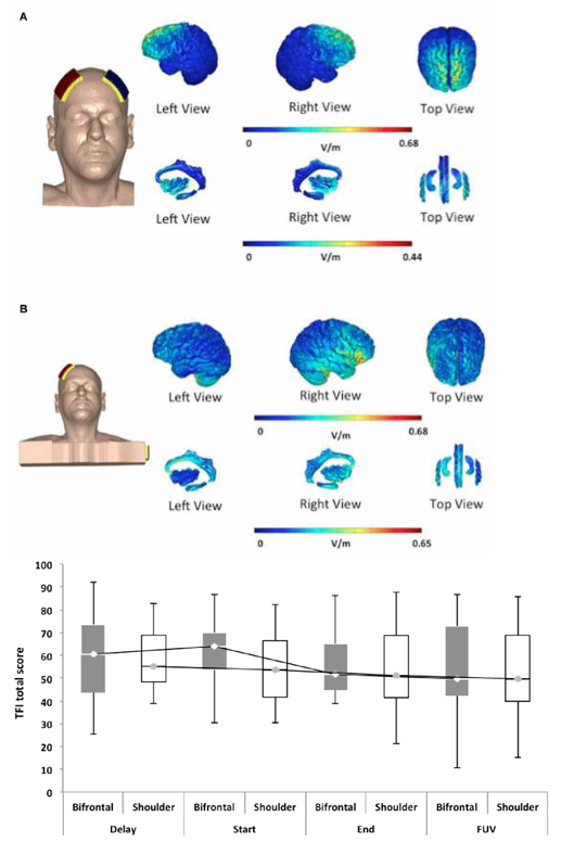
New Paper: Comparing Long-Term Effect of Positioning the Cathode in tDCS in Tinnitus Patients.

Clinical Trial.

Comparison of the Long-Term Effect of Positioning the Cathode in tDCS in Tinnitus Patients. 

Front. Aging Neurosci.  2017, July; 9(217)  doi: 10.3389/fnagi.2017.00217     Download PDF: Comparing long-term effect

Sarah Rabau, Giriraj S. Shekhawat, Mohamed Aboseria, Daniel Griepp, Vincent Van Rompaey,  Marom Bikson6 and Paul Van de Heyning.

Abstract:

Objective: Transcranial direct current stimulation (tDCS) is one of the methods described in the literature to decrease the perceived loudness and distress caused by tinnitus. However, the main effect is not clear and the number of responders to the treatment is variable. The objective of the present study was to investigate the effect of the placement of the cathode on the outcome measurements.

Methods: Patients considered for the trial were chronic non-pulsatile tinnitus patients with complaints for more than 3 months and a Tinnitus Functional Index (TFI) score that exceeded 25. The anode was placed on the right dorsolateral prefrontal cortex (DLPFC). In the first group—“bifrontal”—the cathode was placed on the left DLPFC, while in the second group—“shoulder”—the cathode was placed on the shoulder. Each patient received two sessions of tDCS weekly and eight sessions in total. Evaluations took place on the first visit for an ENT consultation, at the start of therapy, after eight sessions of tDCS and at the follow-up visit, which took place 84 days after the start of the therapy. Subjective outcome measures such as TFI, Visual Analog Scales (VAS) for loudness and percentage of consciousness of tinnitus were administered in every patient.

Results: There was no difference in the results for tinnitus loudness and the distress experienced between the placement of the cathode on the left DLPFC or on the shoulder. In addition, no statistically significant overall effect was found between the four test points. However, up to 39.1% of the patients experienced a decrease in loudness, measured by the VAS for loudness. Moreover, 72% of those in the bifrontal group, but only 46.2% of those in the shoulder group reported some improvement in distress.

Conclusion: While some improvement was noted, this was not statistically significant. Both electrode placements stimulated the right side of the hippocampus, which could be responsible for the effect found in both groups. Further research should rule out the placebo effect and investigate alternative electrode positions.

7-28-17.PNG
Neural Engineering
New Paper: The Influence of Skin Redness on Blinding in Transcranial Direct Current Stimulation Studies: A Crossover Trial

Ezquerro F, Moffa AH, Bikson M, Khadka N, Aparicio LVM, Sampaio BD Jr, Fregni F, Bensenor I,  Lotufo P, Pereira AC, Brunoni AR


Download: PDF Published in Neuromodulation  DOI

Abstract

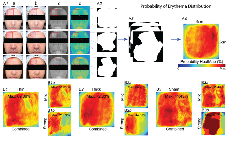
Objective: To evaluate whether and to which extent skin redness (erythema) affects investigator blinding in transcranial direct current stimulation (tDCS) trials.
Material and Methods:Twenty-six volunteers received sham and active tDCS, which was applied with saline-soaked sponges of different thicknesses. High-resolution skin images, taken before and 5, 15, and 30 min after stimulation, were randomized and presented to experienced raters who evaluated erythema intensity and judged on the likelihood of stimulation condition (sham vs. active). In addition, semi-automated image processing generated probability heatmaps and surface area coverage of erythema. Adverse events were also collected.
ResultsErythema was present, but less intense in sham compared to active groups. Erythema intensity was inversely and directly associated to correct sham and active stimulation group allocation, respectively. Our image analyses found that erythema also occurs after sham and its distribution is homogenous below electrodes. Tingling frequency was higher using thin compared to thick sponges, whereas erythema was more intense under thick sponges.
Conclusions:Optimal investigator blinding is achieved when erythema after tDCS is mild. Erythema distribution under the electrode is patchy, occurs after sham tDCS and varies according to sponge thickness. We discuss methods to address skin erythema-related tDCS unblinding.

7-11-17.PNG
Neural Engineering
New Paper: Neuromodulation of Axons

Neuromodulation of Axon Terminals

Cerebral Cortex, 2017; 1–9 doi: 10.1093/cercor/bhx158   Download PDF:NeuromodulationofAxons

Darpan Chakraborty, Dennis Q. Truong, Marom Bikson and Hanoch Kaphzan


Abstract: Understanding which cellular compartments are influenced during neuromodulation underpins any rational effort to explain and optimize outcomes. Axon terminals have long been speculated to be sensitive to polarization, but experimentally informed models for CNS stimulation are lacking. We conducted simultaneous intracellular recording from the neuron soma and axon terminal (blebs) during extracellular stimulation with weak sustained (DC) uniform electric fields in mouse cortical slices. Use of weak direct current stimulation (DCS) allowed isolation and quantification of changes in axon terminal biophysics, relevant to both suprathreshold (e.g., deep brain stimulation, spinal cord stimulation, and transcranial magnetic stimulation) and subthreshold (e.g., transcranial DCS and transcranial alternating current stimulation) neuromodulation approaches. Axon terminals polarized with sensitivity (mV of membrane polarization per V/ m electric field) 4 times than somas. Even weak polarization (<2 mV) of axon terminals significantly changes action potential dynamics (including amplitude, duration, conduction velocity) in response to an intracellular pulse. Regarding a cellular theory of neuromodulation, we explain how suprathreshold CNS stimulation activates the action potential at terminals while subthreshold approaches modulate synaptic efficacy through axon terminal polarization. We demonstrate that by virtue of axon polarization and resulting changes in action potential dynamics, neuromodulation can influence analog– digital information processing.

7-5-17.PNG
Neural Engineering
Neural Engineering Seminar: Laurent Koessler on EEG source localization

Friday 6/23 at 3 pm in CDI 3rd floor conference room (3.352)

Laurent Koessler from CNRS and Lorraine University will be speaking

Title:  Brain source detection and localization using multi-scale EEG recording.

Abstract: In drug-resistant epilepsy surgery investigations, epileptogenic zone and brain functional areas localization are required. This localization relies on scalp and intracerebral EEG recordings. In Nancy (France) I developed a program concerning simultaneous scalp and intracerebral EEG recordings. Using this methodological approach, 1) in vivo human brain tissue conductivities can be estimated, 2) relationship from brain sources to scalp EEG correlates can be studied and 3) non invasive electrical source localization can be validated.


Neural Engineering
Neural Engineering Seminar: Dr. Bashar Badran, June 21

Tomorrow June 21st at 2pm in CDI 3rd floor conference room (3.352)

Bashar Badran from the Medical Universty of South Carolina and University of New Mexico will be speaking

Title: Development, optimization, and neurophysiological effects of transcutaneous auricular vagus nerve stimulation (taVNS)

Abstract: taVNS is an emerging new form of neuromodulation involving transcutaneous electrical stimulation of the auricular branch of the vagus nerve. Still in its infancy and showing much clinical promise, the optimal human stimulation parameters and direct brain effects are undetermined. This lecture will present the findings of two important studies that aim to solve the taVNS problem of infinite parametric solutions. The first, a taVNS parametric study exploring 9 different combinations of pulse width and frequency and their activation of the vagal tone as measured by physiological recordings. The second is a novel multi-modal imaging study that establishes concurrent taVNS/fMRI and explores the direct brain effect of taVNS on the human brain’s BOLD response. These findings aim to establish an aim and direction of the optimal taVNS parameters to guide future trials.

Neural Engineering
New Paper: "Perfect" EEG guided tES and tDCS

Neuroimage. 2017 May 31;157:69-80. doi: 10.1016/j.neuroimage.2017.05.059.

Optimal use of EEG recordings to target active brain areas with transcranial electrical stimulation.

Dmochowski JP, Koessler L, Norcia AM, Bikson M, Parra LC.

Full paper PDF

Abstract: To demonstrate causal relationships between brain and behavior, investigators would like to guide brain stimulation using measurements of neural activity. Particularly promising in this context are electroencephalography (EEG) and transcranial electrical stimulation (TES), as they are linked by a reciprocity principle which, despite being known for decades, has not led to a formalism for relating EEG recordings to optimal stimulation parameters. Here we derive a closed-form expression for the TES configuration that optimally stimulates (i.e., targets) the sources of recorded EEG, without making assumptions about source location or distribution. We also derive a duality between TES targeting and EEG source localization, and demonstrate that in cases where source localization fails, so does the proposed targeting. Numerical simulations with multiple head models confirm these theoretical predictions and quantify the achieved stimulation in terms of focality and intensity. We show that constraining the stimulation currents automatically selects optimal montages that involve only a few (4-7) electrodes, with only incremental loss in performance when targeting focal activations. The proposed technique allows brain scientists and clinicians to rationally target the sources of observed EEG and thus overcomes a major obstacle to the realization of individualized or closed-loop brain stimulation.

6-15-17.PNG
Neural Engineering
Dmochowski and Bikson: Perspective on "IF" Stimulation in Cell Press

“Noninvasive Neuromodulation Goes Deep” Jacek Dmochowski and Marom Bikson, Cell.http://dx.doi.org/10.1016/j.cell.2017.05.017

Modulating deep regions of the brain with noninvasive technology has challenged researchers for decades. In a new study, Grossman et al. leverage the emergence of a slowly oscillating ‘‘beat’’ from intersecting high-frequency electric fields to stimulate deep brain regions, opening a frontier in the biophysics and technology of brain stimulation. Download PDF: FullPaper

6-1-17.PNG
Neural Engineering